This is your brain on whisky
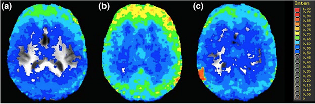
In these brain scans, blue represents a more chaotic brain, while yellow shows less chaos. These images show a person's brain before drinking 6 ounces of whisky (left), immediately after drinking the whisky (middle) and 90 mins after drinking (right).
Inspiration came to engineering professor Mike Noseworthy while listening to the radio.
The medical imaging expert heard a story about possible new penalties for those caught driving with a blood alcohol concentration of .04, or 40 milligrams of alcohol in 100 milliliters of blood.
In Canada, the Criminal Code limit is .08, but several provinces and territories have laws for those found with lesser concentrations of alcohol in their blood.
This got Noseworthy, who has spent 26 years using MRIs to study a variety of medical conditions, thinking about how the brain functions when someone is impaired. Do blood alcohol concentration limits accurately reflect what’s going on in the brain?
“You can’t exactly take a brain biopsy to find out what the alcohol content in the brain is,” said Noseworthy, co-director of McMaster’s School of Biomedical Engineering. “We’re inferring that if it’s in the blood, the same concentration is in the brain.”
Join Mike Noseworthy for an “Ask Me Anything” Q & A on Reddit, April 13 at 1 p.m.
In 2013, Noseworthy and a team of students set out to study the human brain on alcohol. The team recruited 14 male participants who consumed six ounces of whisky each.
Using an MRI scan, the research team looked at particular sections of participants’ brains to extract a nuclear magnetic resonance spectrum, which plots signals of different frequencies that correspond to different molecules.
The researchers then pinpointed how much alcohol was floating through the participants’ craniums.
They also tested how the brain functions while impaired by using blood oxygen level-dependent imaging, a type of MRI method that detects spikes of activity in the brain when bloods starts flowing.
Noseworthy and his team have previously shown that a healthy brain is chaotic, with many networks that act in a difficult-to-predict fashion, allowing it to rapidly adapt to changing circumstances.
When someone’s brain is malfunctioning, as is the case of Alzheimer’s patients, brain patterns are more consistent, making it tough to think clearly and rapidly adapt to change.
With this in mind, Noseworthy and his team sought to determine whether being impaired made the brain less chaotic.
Noseworthy discovered the more intoxicated someone becomes, the less complex their brain patterns become, making it hard to think clearly, respond quickly and react to normal social cues.
While doing an MRI on every impaired driver isn’t feasible, Noseworthy said more research could be done to determine whether a Breathalyzer test produces an accurate reflection of how our brain reacts to alcohol.
“If we could better understand the Breathalyzer test compared to what we’ve found, we may shed some light on what the blood alcohol concentration limit should be,” he says.
Noseworthy’s research appeared in the official journal of the European Society for Magnetic Resonance in Medicine and Biology: Magnetic Resonance Materials in Physics, Biology and Medicine in August with co-authors Noam Soreni and Alexander M. Weber.
